
Fig. 3. Gene deletion of RARα promotes cardiac oxidative stress.
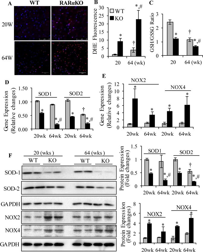
(A) Dihydroethidium (DHE) staining (red) of heart sections collected from WT and RARαKO mice. Scale bars: 50 μM. (B) DHE staining intensity was normalized to section area and plotted. (C) GSH/GSSG ratio. *p<0.05 vs age matched WT; †p<0.05 vs 20 wks WT; #p<0.05 vs 20 wks RARαKO. Cardiac gene expression of SOD1, SOD2 (D) and NOX2 and NOX4 (E). Data are mean value ± SEM (n=6). *p<0.05 vs age matched WT. †p<0.05 vs 20 wks WT; #p<0.05 vs 20 wks RARαKO. (F) Protein expression of SOD1, SOD2, NOX2 and NOX4 was determined and quantified by densitometry. *p<0.05 vs age matched WT; †p<0.05 vs 20 wks WT; #p<0.05 vs 20 wks RARαKO.