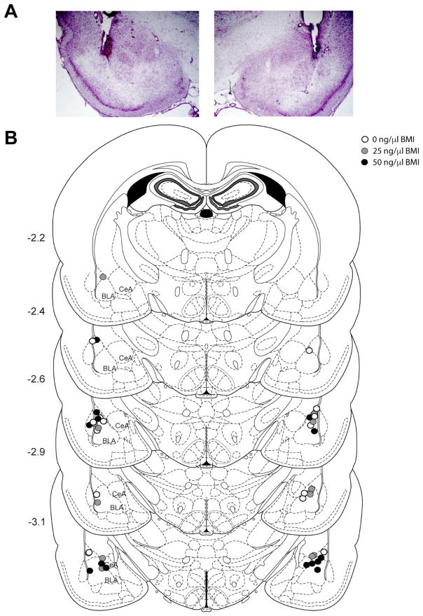
Figure 3.
Basolateral amygdala (BLA) cannulae placements. A) Photograph depicting cannula placements and typical amount of damage caused by the cannulae and obturator/infuser. B) Schematic drawing showing the location of injector tips within the BLA. Rats were excluded (not shown) if their tips were not within or bordering the BLA. Numbers on the left indicate location posterior from bregma. Adapted from Paxinos and Watson (2009). BLA (basolateral amygdala); CeA (central amygdala).

