. 2016 Oct 12;41(4):283–295. doi: 10.5395/rde.2016.41.4.283

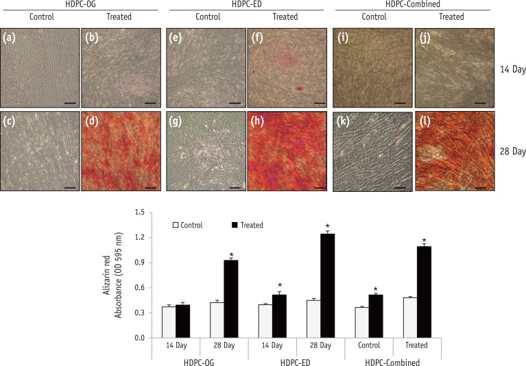
Figure 3. Osteogenic differentiation of HDPCs isolated by three different methods. Alizarin Red S staining of HDPCs-OG (a - d), HDPCs-ED (e - h), and HDPCs-Combined (i - l) cultures at 14 and 28 days after the induction of differentiation. Mineralization progressed at a higher rate in HDPC-ED cultures at 14 days (f) and 28 days (h) after induction than HDPCs-OG (b and d) and HDPCs-Combined (j and l). Scale bar: 50 µm. HDPCs, human dental pulp cells; HDPCs-OG, HDPCs were isolated by the outgrowth method; HDPCs-ED, HDPCs were isolated by the enzymatic digestion method (collagenase/dispase/trypsin); HDPCs-Combined, HDPCs were isolated by the combination of both methods. *Asterisk indicates difference significant between control and treated groups (α = 0.05).

Figure 3