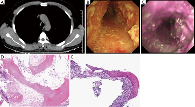
Figure 1.
Imaging and histopathological findings for a 42-year-old man with tracheobronchopathia osteochondroplastica. (A) An axial computed tomography image (soft tissue window) shows multiple calcified nodules in the anterior and lateral walls of the trachea; (B) bronchoscopy shows diffuse white nodules protruding into the tracheal lumen; (C) autofluorescence imaging bronchoscopy shows a magenta color covering the entire circumference of the trachea; (D,E) histopathological examination from biopsy of carina shows submucosal calcification and ossification with fatty bone marrow. (D) (HE; ×40) Mucosal histopathology illustrates squamous metaplasia; (E) (HE; ×100).

