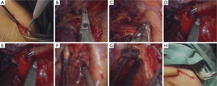
Figure 1.
Single-port VATS in a patient with non-small cell lung cancer. (A) An axillary midline incision (3–5 cm) was made in the fifth intercostal space; (B-E) the right upper pulmonary vein (B), right upper pulmonary artery (C,E) and right upper bronchus (D) were ligated and divided using an endoscopic stapler; (F,G) the resected lymph nodes were of station 2 and 4 (F) and 7 (G); (H) a chest drainage tube was placed through the posterior border of the incision. VATS, video-assisted thoracic surgery.

