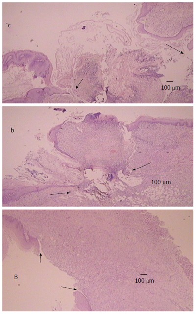
Figure 8.

Illustrative microscopic presentation in rats that underwent esophagogastric anastomosis at post-operative day 4 [controls (c), BPC 157 (b)] and at post-operative day 7 [BPC 157 (B)], hematoxylin eosin staining. Tissue destruction at the anastomosis edges (arrows) were consistently noted in controls (c), which is a presentation that precedes lethal outcome. Initial organization of the exudate (b) (arrows) and wound completely closed with granulation tissue (B) (arrows), which is consistently noted in rats that underwent esophagogastric anastomosis and were treated with BPC 157.
