
Figure 4.
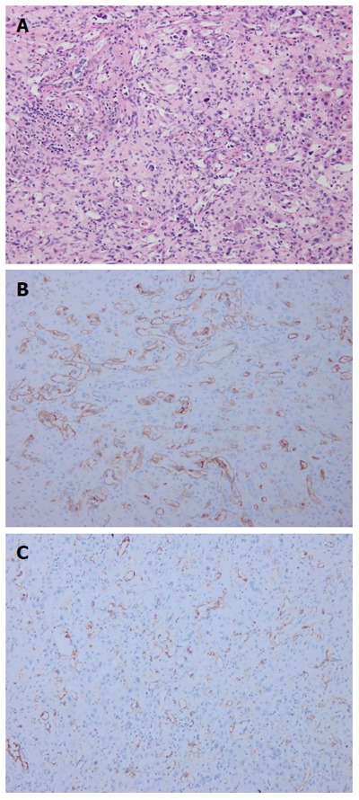
Pathological findings. Pathological investigations identified hepatic epithelioid hemangioendothelioma: A: Hematoxylin-eosin staining revealed abnormal hyperplasia of fibrous tissue combined with vessel-like formations, with scattered neoplastic epithelioid cells; B: Immunohistochemistry showed that the tumor was positive for CD34; C: Immunohistochemistry showed that the tumor was positive for CD31.