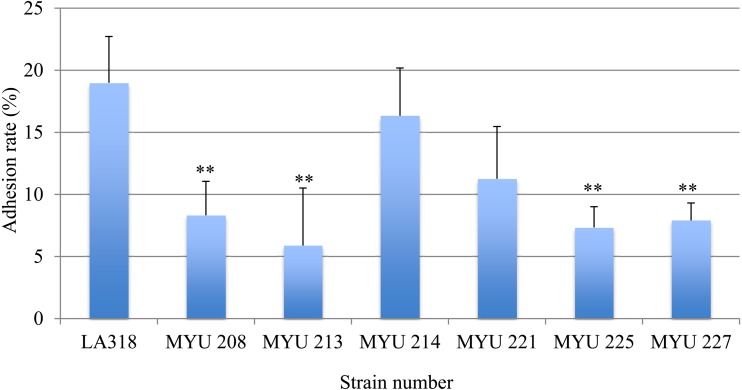
Fig. 2.
Test of adhesion of selected LAB to porcine intestinal mucin (PIM).
Purified unlabeled PIM in PBS was added to each well and incubated overnight at 4°C. After blocking and washing, 100 µL of 1 × 10⁸ cells/ml CFDA-labeled LAB cells in distilled water was added to the wells. The microbial cells were allowed to adhere for 1 hr at 37°C, and the wells were washed three times with PBS to remove non-adherent cells. The cells bound to PIM were released and lysed using 1% SDS/0.1 M NaOH solution and incubated for 1 hr at 60°C. After incubation, the fluorescence intensity (excitation, 485 nm; emission, 538 nm) of the lytic solution was measured. PBS was used as the control in place of PIM. The adhesion value was defined as the value of the mucin-LAB immobilized on the plate reduced by the control value. The results are shown as adhesion rates (%). L. plantarum LA 318 was used as a positive control.
** Significantly different compared with the control strain (LA 318) (p<0.01).

