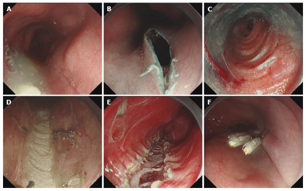
Figure 1.

Case illustration of peroral endoscopic circular myotomy. A: Endoscopy showing dilated esophagus; B: Longitudinal mucosal incision was made to create a tunnel entry; C: Submucosal tunnel; D: Endoscopic image of the circular muscle; E. Circular myotomy; F: Tunnel entry was closed with several clips.
