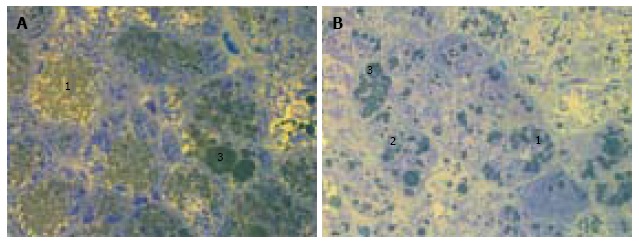
Figure 1.

Liver histology in intact db/db mice. A, B: Microvesicular (1) and mediovesicular (2) lipid accumulation, sporadic large lipid droplets in hepatocytes (3). Light microscopy with yellow filter of semi-thin sections stained with toluidine blue; magnification × 1000.
