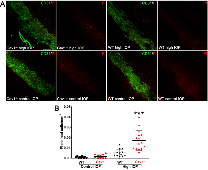
Figure 4. Caveolae protect SC endothelial membranes from IOP-induced damage.
(A) Representative maximum projections of z-stacks from anterior segment wholemounts that encompass the entire SC diameter from Cav-1−/− and littermate controls subjected to IOP elevation (upper panels) or maintained at standing IOP (lower panels). The SC endothelium was co-stained with CD31 (green) and PI-stained nuclei are labeled in red. Scale bar = 100 μm. (B) Quantification of PI-stained nuclei in each image stack (from n = 5 mice/genotype). For statistical analysis, multiple images from each eye were averaged to yield a single value of PI cells/mm3 per mouse. ***p ≤ 0.001, one-way ANOVA and Newman-Keuls post hoc analysis.

