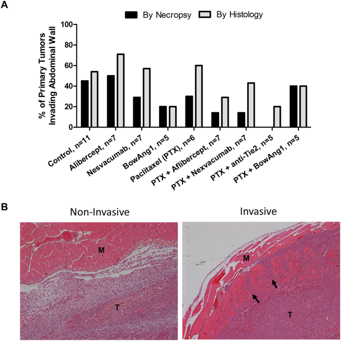
Figure 3. Differential treatment effects on primary breast tumor invasiveness into the abdominal wall.
14 days after orthotopic implantation of 2 × 10⁶ LM2-4 cells, mice bearing ~150-mm3 primary breast tumors were randomized and administered with either the controls (PBS vehicle or IgG1 isotype), aflibercept (anti-VEGF-A/VEGF-B/PlGF), nesvacumab (anti-Ang2), BowAng1, or an anti-Tie2 antibody, with or without paclitaxel chemotherapy, for 2 weeks. On day 29 post-implantation, all mice were sacrificed and their primary breast tumors were examined during necropsy and confirmed histologically for signs of invasion into the abdominal wall. (A) The incidence (%) of invaded tumors per treatment group is plotted. P > 0.05 by Fisher’s exact test. (B) Representative microscopy images from the histological analysis of primary LM2-4 breast tumors for invasions into the adjacent abdominal wall by hematoxylin and eosin staining. “M” denotes abdominal wall muscle. “T” denotes tumor cells. Black arrows mark regions where tumor cells are infiltrating into the abdominal wall and separating muscular fascicles.

