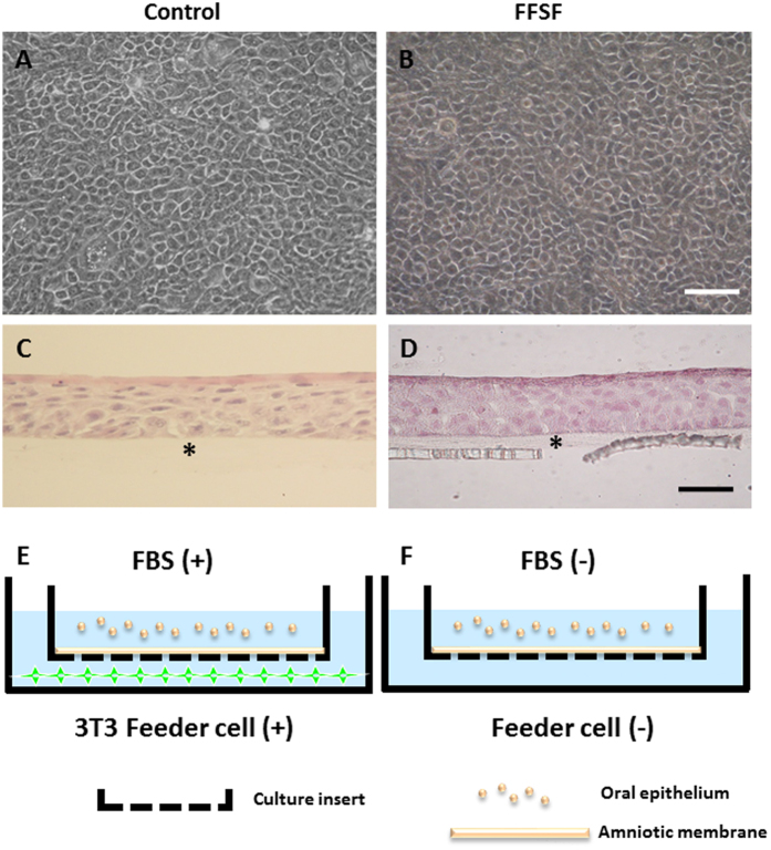
Figure 1. Representative phase contrast images and histological examinations of the cultivated oral mucosal epithelial cell sheet (COMECS).
Phase contrast pictures showing confluent primary culture of oral mucosal epithelial cells in Control (A) and FFSF (B) after 7 days in culture; light micrographs showing cross-sections of COMECS in Control (C) and FFSF (D) culture system stained with hematoxylin and eosin; illustration of Control (E) and FFSF (F) culture conditions. Asterisks indicate denuded amniotic membrane. Scale bars = 100 μm.

