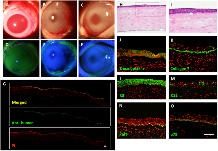
Figure 6. Xeno-transplantation of cultivated oral mucosal epithelial cell sheet (COMECS).
Representative slit-lamp pictures of a rabbit taken just before surgery, with and without fluorescein (A,D) 7 days post transplantation, with and without fluorescein (B,E) and 14-days post transplantation, with and without fluorescein (C,F); immunofluorescence of anti-human nuclei at the transplanted area (G); representative HE staining (H,I) and immunofluorescence of desmoplakin (J) collagen 7 (K) keratin 3 (L) 12 (M) Ki67 (N) and p75 (O) in the transplanted FFSF COMECS. Nuclei were stained with propidium iodide (red). Scale bars = 100 μm.

