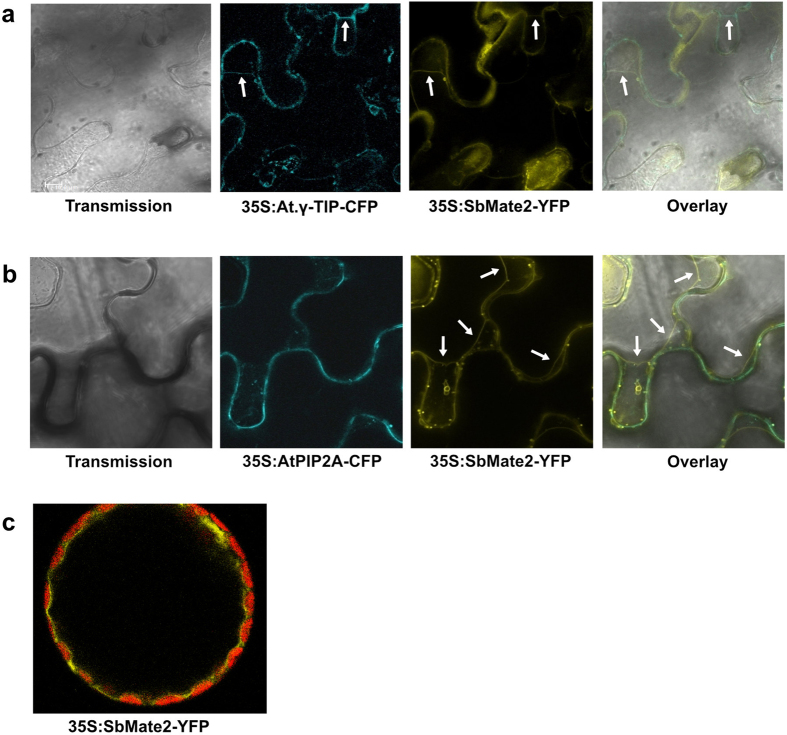
Figure 2. Tonoplast localisation of the SbMATE2-YFP fusion protein in epidermal cells and protoplasts of Nicotiana benthamiana.
A SbMATE2-YFP fusion protein under the control of the 35S-CaMV promoter was transiently expressed in N. benthamiana using Agrobacterium infiltration. The SbMATE2-YFP construct was coinfiltrated with expression constructs for either: (a) At.γ-TIP-CFP, a marker for the vacuolar membrane, or (b) AtPIP2A-CFP, a marker for the plasma membrane38. The localisation of the fusion proteins was visualised using confocal microscopy, and the SbMATE-YFP signal co-localised with that of γ-TIP-CIP, showing a tonoplast localisation. Arrows indicate positions where the tonoplast is not adjacent to the plasma membrane. (c) Localisation of SbMATE2-YFP (in yellow) to the vacuolar membrane in isolated N. benthamiana protoplasts. Chloroplasts situated between the vacuolar and plasma membranes are visualised by their autofluorescence (in red).

