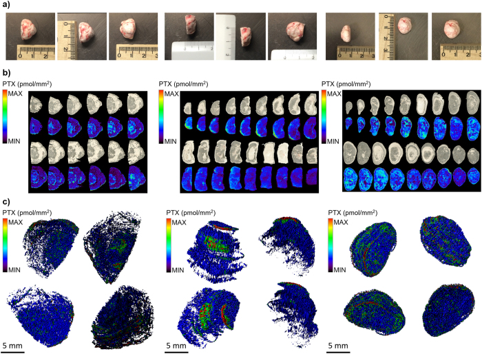
Figure 1. Three-dimensional reconstruction of tumors.
(a) Mesotheliomas and their dimensions. (b) Serial MALDI 2D images of different tumors, compared to optical images of the same slices before acquisition. In each image, colors are normalized to the same scale that appears in different experiments and indicate the same drug concentration. (c) 3D representation of PTX distribution inside MPM487 mesotheliomas.

