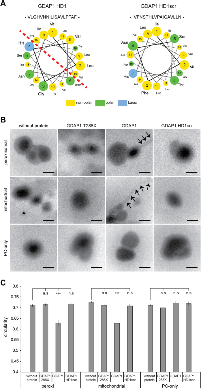
Figure 2. GDAP1 tubulates liposomes dependent on the HD1 and the lipid composition.
(A) Helical wheel representation of residues of the HD1 reveals the amphipathic pattern of hydrophilic and hydrophobic amino acids. This pattern is broken up by scrambling the primary sequence of the HD1 (HD1scr). (B) Electron microscopy of negatively stained liposomes of phosphatidylcholine (PC) or of lipid compositions resembling the mitochondrial outer membrane (Mito) or peroxisomal membrane (Peroxi) were incubated with in vitro translated GDAP1, GDAP1 HD1scr, GDAP1 lacking HD1 and TMD (GDAP1 T288X), or were left untreated. All liposomes have primarily multilamellar appearances. Only the addition of GDAP1 to Mito- and Peroxi-liposomes caused tubulation (arrows). Scale bars: 50 nm. (C) Per preparation 16 to 25 electron micrographs were taken blindly. On electronic pictures all discrete liposomes were selected automatically and the circularity of the objects was determined. The graph depicts the mean and the s.e.m. of all liposomes per condition (n = 79 to 197) from independent preparations, paired t-test ***P-value < 0.0005.

