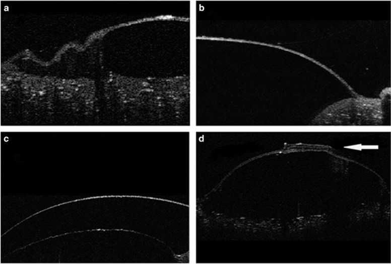
Figure 1.
Topcon OCT: (a) Type-1 BB showing two curvilinear lines. The anterior line represents DL and banded zone of DM. (b) Type- 2 BB showing two curvilinear lines that represent banded and non-banded zones of DM. (c) Mixed BB where the anterior line represents DL and the posterior line represents DM. (d) Type-1 BB from which the DM was partially peeled off. The peeled DM is folded on itself (arrow). The OCT image to the right of the peeled DM is a single line and that to the left has a double-contour as seen in the posterior wall of a type-1 BB.

