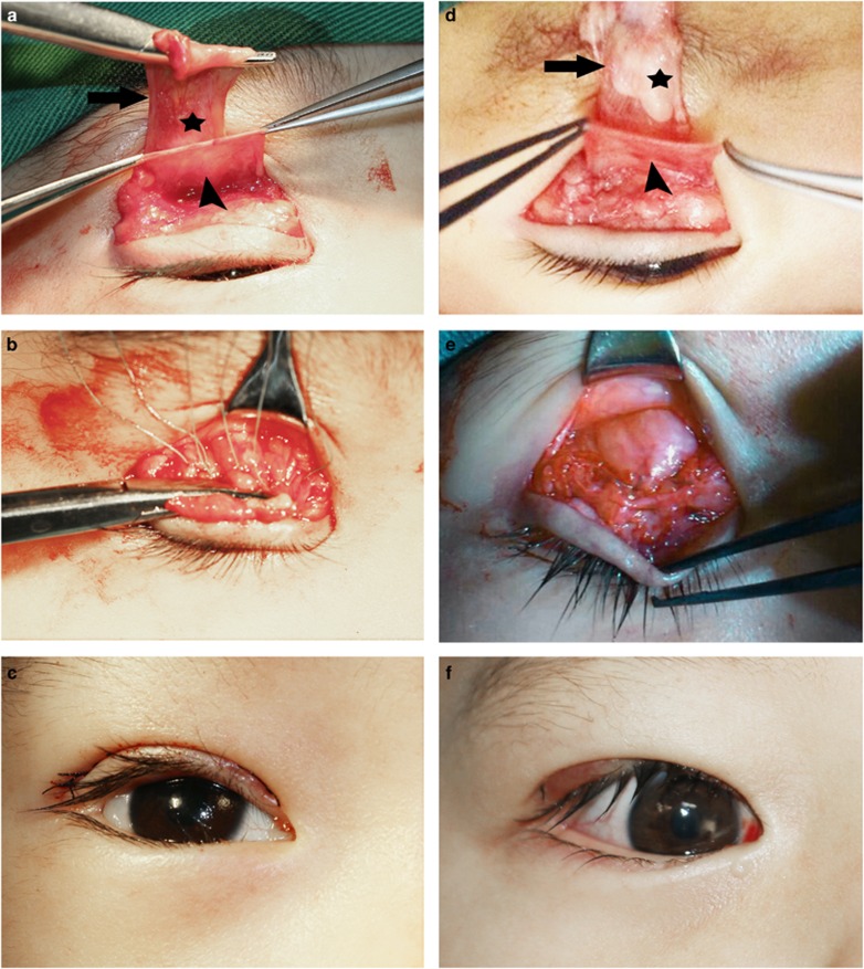
Figure 1.
Photographs of two patients who underwent levator resection and SLSF suspension. (a) Levator aponeurosis was found to be thin and fragile (*). Arrow, levator aponeurosis; arrowhead, SLSF. (d) Fatty degeneration was found in levator aponeurosis (*). Arrow, levator aponeurosis; arrowhead, SLSF. (b, e) Three mattress sutures were placed between the tarsal plate and the levator aponeurosis-SLSF composite flap. (c, f) Postoperative view.

