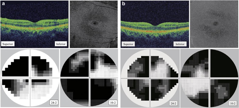
Figure 2.
MME observed in SD-OCT B-scan vertical scan image and en-face imaging. (a) A 57-year-old woman with primary open-angle glaucoma and MME in her right eye (patient 2). (b) A 64-year-old woman with normal tension glaucoma and MME in her left eye (patient 3). MME was commonly observed under the papillomacular nerve fiber bundle. All patients had visual field defects in the central 24 or 10 degrees.

