
Figure 1.
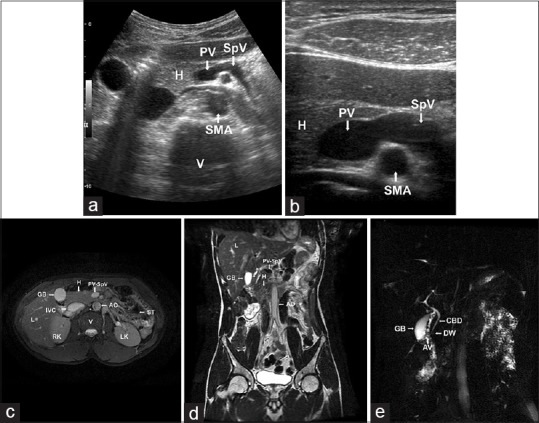
(a and b) Ultrasonographic image in transverse plane showing the head and uncinate process of the pancreas; the neck, body, and tail of the pancreas were absent anterior to portal confluence and splenic vein. H: Head of the pancreas; PV: Portal vein; SpV: Splenic vein; SMA: Superior mesenteric artery; V: Vertebra. (c) Axial T2-weighted magnetic resonance image showing normal head and uncinate process of the pancreas with the absence of neck, body, and tail of the pancreas. (d) Coronal T2-weighted magnetic resonance image showing normal head and uncinate process of the pancreas with the absence of neck, body, and tail of the pancreas. H: Head of the pancreas; PV-SpV: Portal vein-Splenic vein confluence; AO: Abdominal Aorta; IVC: Inferior vena cava; V: Vertebra; RK: Right Kidney; LK: Left Kidney; ST: Air-filled Stomach; L: Liver; GB: Gallbladder. (e) Maximum intensity projection image in magnetic resonance cholangiopancreatography showing common bile duct and duct of Wirsung. The duct of Santorini is not visualized. CBD: Common bile duct; DW: Duct of Wirsung; AV: Ampulla of Vater; GB: Gallbladder