
Figure 1.
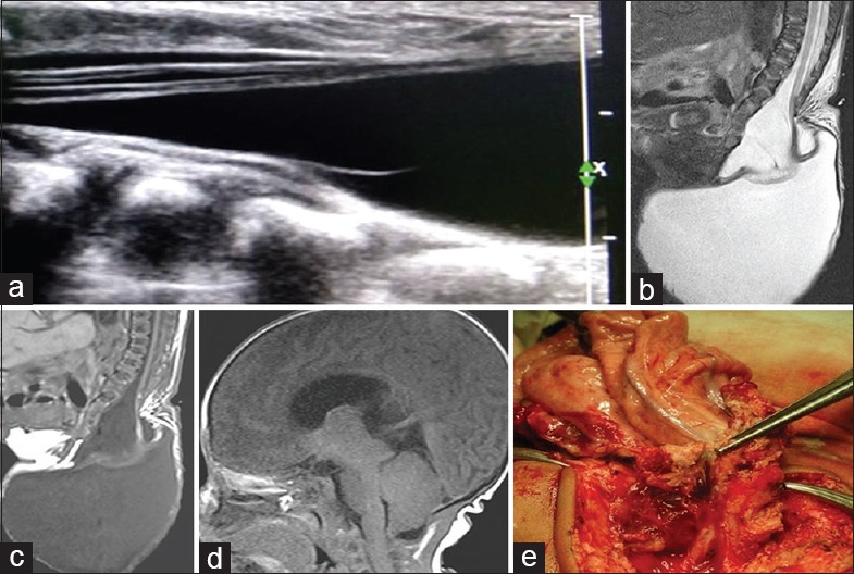
(a) Gray-scale spinal ultrasonography shows herniation of terminal cord and cerebrospinal fluid out of spinal canal with tethering of cord and focal syrinx. (b) Sagittal T2-weighted of L-S spine shows posterior elements defect in the sacral region through which there is herniation of large cerebrospinal fluid intensity cystic lesion in the sacrococcygeal region causing tethering of cord and mild syrinx formation. (c) Sagittal T1-weighted of L-S spine show findings similar to T2-weighted. No evidence of lipomatous tissue is noted. (d) Sagittal T1-weighted of brain shows caudal herniation of cerebellar vermis and brainstem, slit-like fourth ventricle, tectal beaking, and large massa intermedia, suggesting Chiari II malformation. (e) Peroperative photograph shows inner surface of the meningomyelocele sac attached to neural placode