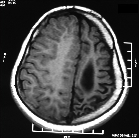
Figure 1.

Magnetic resonance imaging of the brain showing diffuse atrophy of the left cerebral hemisphere with dilatation of the left lateral ventricle and prominence of sulci over the left cerebral hemisphere. There is also compensatory thickening of the skull vault
