Figure 1.

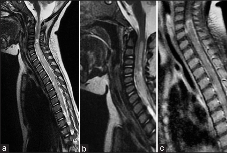
(a and b) Sagittal T2 weighted images showing diffuse hyperintensity in spinal cord extending from c4 (1a) and syrinx above c4 (1b). (c) Sagittal T1 weighted post contrast images showing patchy enhancement in cord

(a and b) Sagittal T2 weighted images showing diffuse hyperintensity in spinal cord extending from c4 (1a) and syrinx above c4 (1b). (c) Sagittal T1 weighted post contrast images showing patchy enhancement in cord