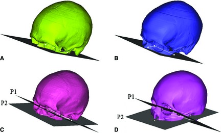
Figure 2.

Four cutting planes utilized for improved image registration. (A) Mid‐orbit: translated below the occipital condyles. (B) Below Orbit: translated below the orbit. (C) Infraorbital Foramen: one plane translated to below the occipital condyles with another at the infraorbital foramen. (D) Nose: one plane translated to below the occipital condyles with another at the inferior nasal bone. (P1 = Plane 1, P2 = Plane 2).
