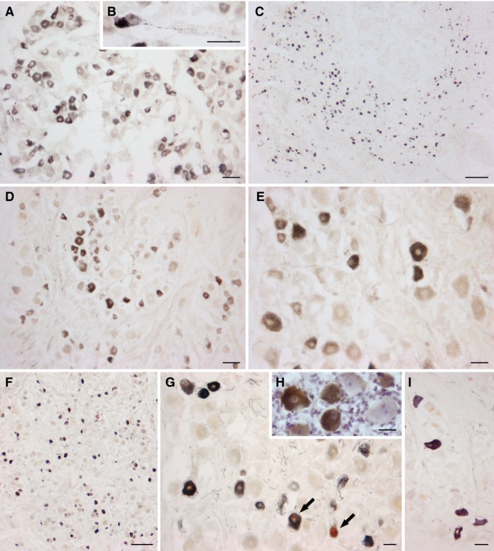
Figure 2.

Human trigeminal ganglion. TRPV1‐LI neurons from two pre‐term newborns (A, B, case 1; C, case 3), a full‐term newborn (D, E, case 5) and two adults (F, G, H, case 8; I, case 10). (B) High magnification of a TRPV1‐labelled TG neuron with a varicose proximal process. (H) Immunostained section with Mayer's modified haematoxylin counterstaining. Arrows in (G) point to neurons containing small (right) and large (left) light brown deposits of lipofuscins. Scale bars: (A, D, E, G, I) 50 μm, (C, F) 250 μm, (B, H) 25 μm.
