Figure 2.
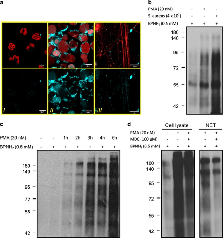
Exogenous monoamines mimic endogenous polyamine incorporation occurring in NETosing neutrophils. (a) Representative images of BPNH2 incorporation into neutrophil intracellular and NET proteins upon 4-h activation with PMA. Neutrophils from healthy donors were preincubated with BPNH2 for 1 h and then stimulated with PMA or left unstimulated for 4 h (control). (I) Shows BPNH2 incorporation in resting neutrophils; (II) shows BPNH2 incorporation in activated neutrophils; and (III) shows BPNH2 incorporation into NET proteins. Upper panels show BPNH2 (light blue) and DNA (red); lower panels show BPNH2 staining alone. The experiment was repeated at least three times with neutrophils from independent healthy donors, with similar results. The original magnification was × 60, the scale bar represents 10 μm. (b) Detection of exogenous monoamine (BPNH2) incorporation into cellular proteins in activated neutrophils by western blot. Neutrophils from a healthy donor were preincubated with BPNH2 and then stimulated either with PMA or S. aureus or left unstimulated for 4 h (control). Levels of BPNH2 incorporation into cellular proteins were determined from cell lysate with western blot. (c) Time-dependent incorporation of BPNH2 into neutrophil cellular proteins upon activation. Neutrophils from a healthy donor were preincubated with BPNH2 and then stimulated with PMA for 0–5 h. BPNH2 incorporation levels were detected as in Figure 2b. (d) Effect of MDC on BPNH2 incorporation into cellular and NET proteins. Neutrophils were preincubated with BPNH2 or BPNH2 and MDC followed by PMA stimulation for 4 h or left unstimulated (control). Cellular and NET proteins were isolated and BPNH2 incorporation levels were detected as in Figure 2b

