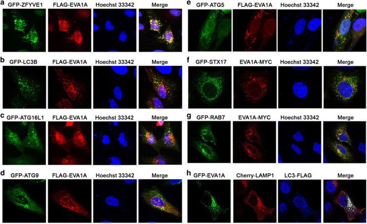
Figure 3.
EVA1A colocalizes with the autophagosomal membrane. Confocal microscopy images are shown in U2OS cells: co-transfected with FLAG-EVA1A (or MYC-EVA1A) and GFP-ZFYVE1 (a), GFP–LC3B (b), GFP–ATG16L1 (c), GFP-ATG9 (d), GFP-ATG5 (e), GFP-STX17 (f) or GFP-RAB7 (g) and then immunostained with an anti-FLAG (or MYC) antibody after 24 h. Nuclei were stained with Hoechst 33342. (h) Co-transfected with GFP-EVA1A, Cherry-LAMP1 and LC3B-FLAG, and then immunostained with an anti-FLAG antibody after 24 h

