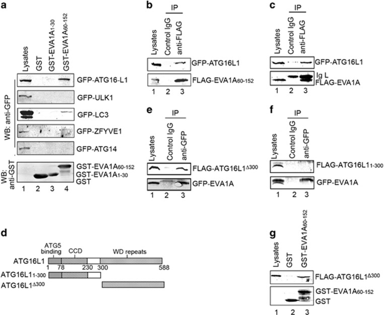
Figure 7.
EVA1A is associated with ATG16L1 via its C-terminal. (a) GST-EVA1A1–30, the GST-EVA1A60–152 fusion protein, and the GST protein immobilized on glutathione-sepharose beads were incubated with HeLa cell lysates containing GFP-DFCP1, GFP-ULK1, GFP-LC3, GFP-ATG14 or GFP–ATG16L1, respectively. GFP and GST were detected in the washed beads by western blot. (b and c) HeLa cells were co-transfected with GFP–ATG16L1 and FLAG-EVA1A60–152 or FLAG-EVA1A for 24 h. Total cell extracts were subjected to IP using either an anti-FLAG or a nonspecific control mIgG as indicated; GFP and FLAG were detected in the washed beads by western blot. (d) Schematic representations of the WT ATG16L1 and its mutants. (e and f) HeLa cells were co-transfected with GFP-EVA1A and FLAG-ATG16L1△300 or FLAG-ATG16L11–300 for 24 h. Total cell extracts were subjected to IP using either an anti-FLAG or a nonspecific control mIgG, as indicated. GFP and FLAG were detected in the washed beads by western blot. (g) GST-EVA1A60–152 fusion protein and the GST protein immobilized on glutathione-sepharose beads were incubated with FLAG-ATG16L1△300 transfected HeLa cell lysates at 4 °C for 4 h. GFP and GST were detected in the washed beads by western blot

