Figure 8.
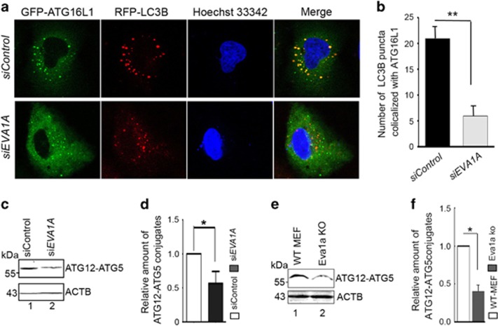
Knockdown of EVA1A decreases the colocalization between LC3B and ATG16L1. (a) U2OS cells were transfected with siControl or siEVA1A for 24 h, transfected with GFP–ATG16L1 and RFP-LC3B for 24 h, and treated with EBSS for the last 2 h. Representative confocal microscopy images were shown. (b) Cell treatment was as same as (a). The number of RFP-LC3B puncta that colocalize with GFP–ATG16L1 were analyzed. Data are the mean±S.D. of at least 50 cells scored. **P<0.01. (c) Western blot analysis of ATG12–ATG5 conjugates in U2OS cells transfected with siControl or siEVA1A for 48 h. (d) Quantification of the amount of ATG12–ATG5 conjugates relative to ACTB. The average value in the siControl treated cells was normalized to 1. Data are the mean±S.D. of results from three experiments.*P<0.05. (e) Western blot analysis of ATG12–ATG5 conjugates in MEFs treated as indicated. (f) Quantification of the amounts of ATG12–ATG5 conjugates relative to ACTB. The average value in WT MEFs was normalized to 1. Data are expressed as the means±S.D. of the results from three experiments. *P<0.05, ** P<0.01

