Figure 1.
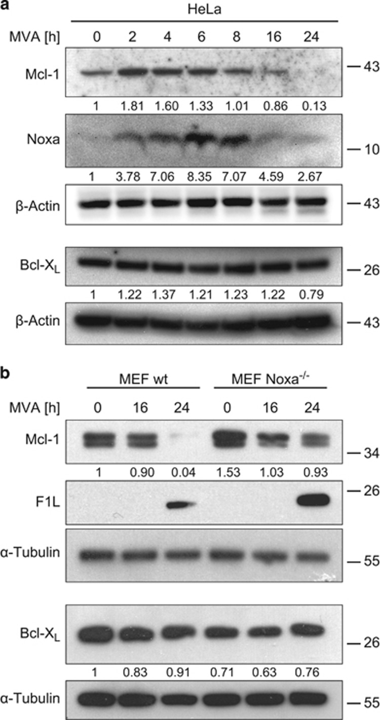
(a) Infection of HeLa cells with MVA leads to the upregulation of Noxa and the loss of Mcl-1 protein. HeLa cells were infected with MVA (MOI=10) for the times indicated. Protein levels were determined by western blotting. Signals were quantified by densitometry and changes in Mcl-1, Noxa or Bcl-XL/β-Actin ratios normalized to the uninfected control are shown below the blots. Data are representative of at least four independent experiments. (b) MVA infection leads to a reduction in Mcl-1 levels but has no effect on Bcl-XL levels in MEFs. MEF wt or Noxa−/− cells were infected with MVA (MOI=10) for 16 or 24 h. Protein levels were assessed by western blotting. Signals were quantified by densitometry and changes in Mcl-1 or Bcl-XL/α-Tubulin ratios normalized to the uninfected wt control are shown below the blots. Data are representative of at least six independent experiments

