Figure 1.
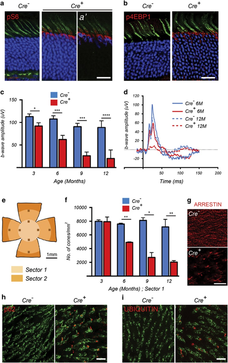
Loss of Tsc1 in cones of wild-type mice leads to defective autophagy and progressive decline in cone function. Data shown are from mice harboring the Tsc1c/c allele. (a and b) Immunofluorescence analyses on retinal cryosections for phosphorylated S6 (a) and 4EBP1 (b) (red signal) at 2 months of age. Cones were detected by PNA staining (green signal). Blue indicates nuclear DAPI, except in (a′), where it indicates expression of the CRE recombinase in cones. (c) Evaluation of cone function over time showing average b-wave amplitudes and representative photopic ERG traces (d) at indicated time points. Data are representative of recordings from at least six mice per genotype (*P<0.05, ***P<0.001, ****P<0.0001 by Student's t-test). (e–g) Evaluation of cone survival: schematic of retinal flat mount (e) indicating the two sectors (radii: 1 and 2mm) that were used to count cones. (f) Bar graphs representing the average number of cone arrestin-positive cones per mm2 in Sector 1 over time. Data are representative of at least two mice in each group (*P<0.05, **P<0.01 by Student's t-test). (g) Representative examples of cone arrestin staining (red signal) from a region in Sector 1 in Cre− and Cre+ mice at 12 months of age. (h) Immunofluorescence analysis on retinal flat mounts (red signal) for p62 and UBIQUITIN (i) at 2 months of age. Cones are marked in green by PNA. Scale bars: 20 μm

