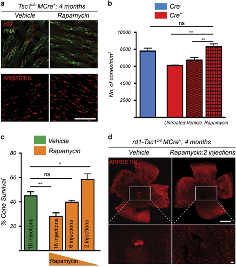
Figure 5.
Rapamycin reverses the autophagy defect and improves cone survival upon Tsc1 loss. (a) Immunofluorescence analysis on retinal flat mounts from Tsc1c/cCre+ mice showing clearance of p62 aggregates in cones (upper panel, red signal; green: PNA) and recovery of cone arrestin (lower panel, red signal) expression upon delivery of rapamycin between P28 to 4 months of age. Images were acquired at 1mm radius from the optic nerve (Sector 1, see Figure 1e). Scale bars: 20 μm. (b) Quantification of cone arrestin-positive cones in Sector 1 at 4 months of age of Tsc1c/cCre+ mice injected with rapamycin or vehicle from P28 onwards (18 injections). Data are representative of at least three mice in each case. **P<0.01 by Student's t-test. (c) Quantification of cone survival in rd1-Tsc1c/cCre+ mice at 4 months of age upon injection with vehicle or rapamycin starting at P28. Number of rapamycin injections is indicated in bar. Data are representative of at least six mice in each group (*P<0.05, **P<0.01 by Student's t-test). (d) Representative retinal flat mounts of rd1-Tsc1c/cCre+ mice at 4 months of age showing more central cones when two injections of rapamycin were administered (red signal: cone arrestin; scale bars: 1 mm)

