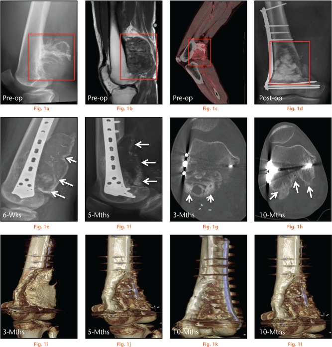
Time course observations of bone defect from a wide periosteal chondrosarcoma resection, treated with hydroxyapatite-calcium sulphate (HA-CS) and HA-CS-gentamicin (G) biomaterial. Panels (a), (b) and (c) represent pre-operative radiograph, MRI and PET-CT images of the tumour (red box), while panel (d) shows the bone defect filled with the biomaterial (red box) post-operatively. Arrows in the radiographs in panel (e) indicate bone formation in areas of direct muscle contact with HA-CS and HA-CS-G six weeks post-operatively, while arrows in panel (f) indicate reduced opacity of the regenerate at five months. Arrows in CT images in panels (g) and (h), respectively, indicate bone formation in the treated area posterior to the distal supracondylar region at three months post-operatively, progressively remodeling at ten months. Panels (i) through to (l) represent 3D reconstructions of CT images at three (i), five (j) and ten months (k), (l) showing progressive bone remodeling around the defect.

An official website of the United States government
Here's how you know
Official websites use .gov
A
.gov website belongs to an official
government organization in the United States.
Secure .gov websites use HTTPS
A lock (
) or https:// means you've safely
connected to the .gov website. Share sensitive
information only on official, secure websites.
