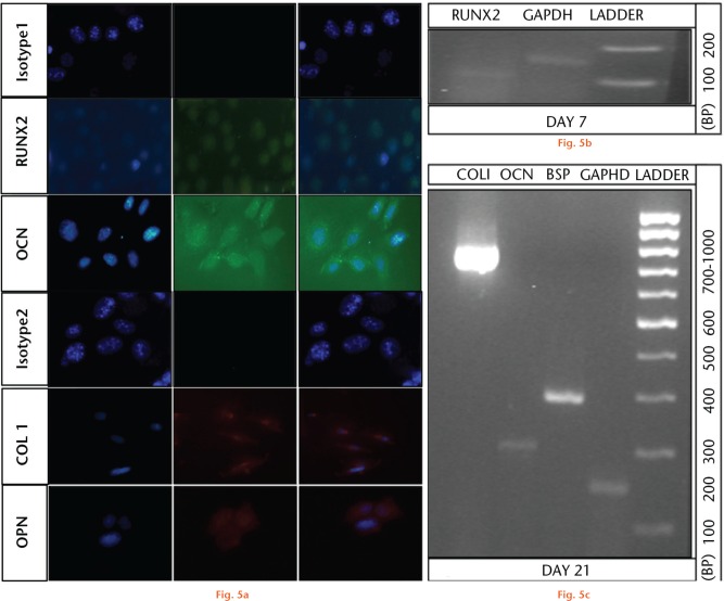
Immunocytochemical and real-time polymerase chain reaction analysis of C2C12 muscle myoblasts seeded on hydroxyapatite-calcium sulphate (HA-CS) biomaterial. C2C12 cells seeded on HA-CS were analysed using immunocytochemistry to visualise osteogenic differentiation. Cells were stained after a period of seven and 21 days post-seeding. The first row (a) shows the isotype control for goat anti-rabbit, Alexa Fluor 488 while the fourth row shows the isotype control for goat anti-mouse Cy3 secondary antibodies. Cells stained positive for osteogenic markers like RUNX2 (second row) at day seven, and polyclonal OCN (third row), Col I (fifth row), and monoclonal OPN (sixth row), after 21 days post-seeding. Images in the left hand panels (all rows) indicate nuclear staining using 4’,6-diamidino-2-phenylindole staining while images in the middle panels (all rows) indicate antibody-based detection of respective target proteins while right hand panels (all rows) depict respective merged images (all 50 μm). Early onset of osteogenic differentiation was confirmed by the presence of the RUNX2 gene in C2C12 cells seeded on HA-CS after seven days (b). Osteoblastic maturation (21 days post-seeding) of muscle cells was confirmed by the presence of osteoblastic gene coding for Col I (lane 1), OCN (lane 2), polyclonal bone sialoprotein (lane 3), and housekeeping gene glyceraldehyde 3-phosphate dehydrogenase (lane 4) with control ladder in (c), lane 5.

An official website of the United States government
Here's how you know
Official websites use .gov
A
.gov website belongs to an official
government organization in the United States.
Secure .gov websites use HTTPS
A lock (
) or https:// means you've safely
connected to the .gov website. Share sensitive
information only on official, secure websites.
