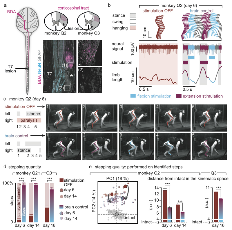
Figure 4. The brain-spinal interface alleviates gait deficits after spinal cord injury.
(a) Diagram illustrating the location of the lesion and corticospinal tract labelling using biotinylated dextran amine (BDA). Right, anatomical reconstructions of spinal segments containing the lesion (grey), for monkeys Q2 and Q3. Photographs including insets, showing a longitudinal view of the lesioned spinal cord wherein astrocytes (GFAP, grey), neurons (NeuN, cyan) and corticospinal tract axons (BDA, pink) are labelled. Asterisk, lesion. Scale bars, overview: 500µm. Insets: 50µm. (b) Gait cycles performed during locomotion without stimulation and during brain-controlled stimulation of both flexion and extension hotspots in monkey Q2 at 6 days post-lesion. Conventions, same as Figure 3. Limb paralysis in red. (c) Snapshots extracted from video recordings showing a sequence of leg movements without stimulation and during brain-controlled stimulation (monkey Q2, 6 days post-injury). Timeline indicates video snapshot timing. Legend refers to panels d and e. (d) Bar plots reporting the ratio between of steps performed by the affected versus unaffected leg by each monkey without stimulation (n = 6 for day 6 and n = 39 for day 14 for Q2, n = 68 for Q3) and during brain-controlled stimulation (n = 12 for day 6 and n = 93 for day 14 for Q2, n = 31 for Q3). (e) Principal component (PC) analysis applied on 26 gait parameters for Q2. All the gait cycles corresponding to limb paralysis or stumbling have been excluded from this analysis. Each gait cycle is shown in the space defined by PC1 and PC2. Bar plots reporting the mean Euclidean distance between pre-lesion and post-lesion gait cycles corresponding to steps, calculated in the entire kinematic space. **,*** significant difference at p < 0.01 and p < 0.001, respectively using bootstrapping (panel d) or Wilcoxon ranksum test (panel e).

