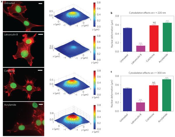
Figure 4. Nanopillar-induced nuclear deformation depends on the integrity of cytoskeletal components.
a, Latrunculin B treatment results in decreased nuclear deformation, whereas acrylamide treatment shows increased nuclear deformation compared with untreated cells. Colchicine did not alter the extent of nuclear deformation. Left column: Fluorescence images showing cell morphology without treatment and under treatment with different cytoskeletal inhibitors: latrunculin B for actin filaments, colchicine for microtubules, and acrylamide for intermediate filaments. Actin was stained with phalloidin-Alexa568 (red) and the nuclear envelope was immunostained for lamin A (green). Scale bars, 10 μm. Right column: Surface plots showing average deformed nuclear surfaces under the different inhibitor treatments. b,c, The change in the depth of nuclear deformation showed the same response to inhibitor treatments on nanopillars with radii of 220 nm (b) and 300 nm (c). All measurements were performed in triplicate. The numbers of nanopillars and nuclei included in each sample are provided in Supplementary Table 1. One-way analysis of variance (ANOVA) was performed on the results in b and c, comparing each group to the untreated cells. NS, no significant difference. ***P < 0.001. Error bars indicate standard error of the mean.

