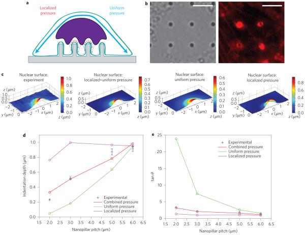
Figure 6. Finite-element analysis suggests that nanopillar-induced deformation is a cumulative result of both the actin cap above the nucleus and actin accumulated around the nanopillar.
a, Illustration of the mechanical model, in which the force pulling the nucleus toward the nanopillars can either be uniformly applied to the apical surface of the nucleus, locally applied around the nanopillar, or a combination of the two. b, Local actin accumulation around nanopillars. Left: Bright-field image showing nanopillar locations. Right: Fluorescence image of actin staining showing bright rings around nanopillars with 300 nm radii. Scale bars, 5 μm. c, Nuclear deformation surfaces display the differential effects of applied pressure profiles. From left to right: experimental surface; simulated surface under combined uniform and localized pressure; simulated surface under uniform pressure only; simulated surface under localized pressure only. d, Of the simulated pressure profiles, the combined uniform and localized pressure most closely reproduces the experimental dependence of nuclear deformation depth on nanopillar pitch. e, Experimental measurements of the deformation angle, shown in blue, match the results from combined pressure more closely than those from either localized or uniform pressure alone.

