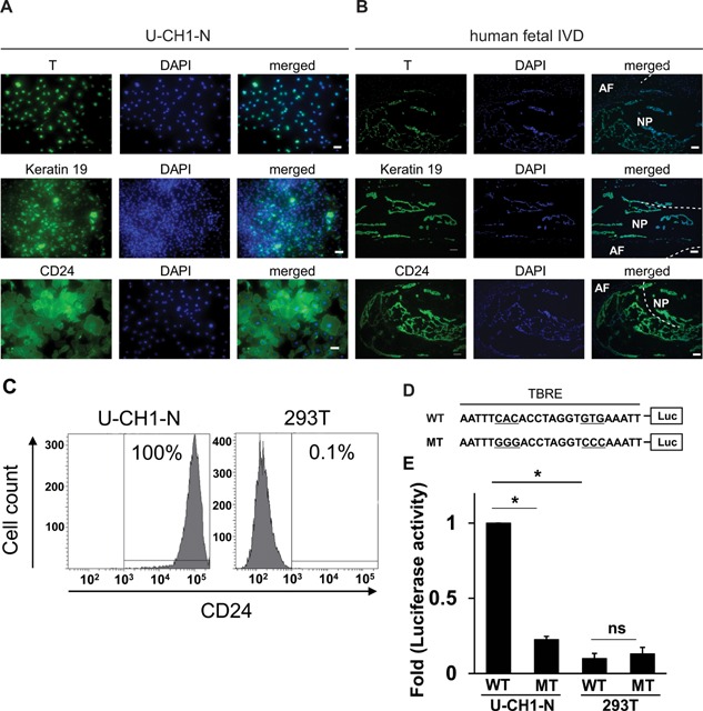
Figure 1.

Expression of the NP markers in U‐CH1‐N cells and human fetal NP cells (NP). (A and B) U‐CH1‐N cells (A) and human fetal IVD (B) were immunostained for T, keratin 19, and CD24, and counterstained with DAPI. Scale bar, 50 µm. (C) Flow cytometric analysis of CD24 expression in U‐CH1‐N cells and 293T cells. (D) A schematic of WT and MT T‐box responsive element (TBRE) constructs used for reporter assay. (E) Luciferase reporter assay using WT and MT reporter constructs. Data are represented as mean ± SEM of three independent experiments performed in triplicate (n = 3). *p < 0.05; ns, not significant.
