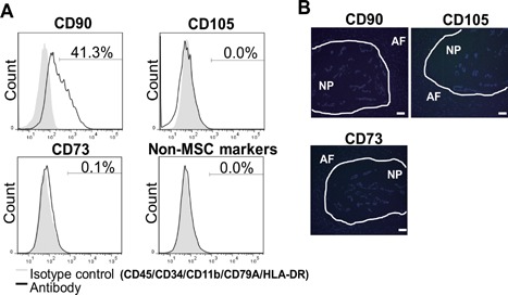
Figure 2.

Analysis of cell surface markers in U‐CH1‐N cells and human fetal NP cells. (A) Flow cytometry analysis of cell surface markers in U‐CH1‐N cells. (B) Human fetal IVD sections immunostained for CD73, CD90, and CD105 and counterstained with DAPI. Scale bar, 50 µm.
