
Figure 3.
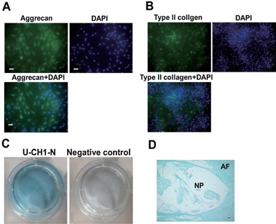
U‐CH1‐N cells exhibit chondrogenic capacity (A) U‐CH1‐N cells immunostained for aggrecan (A) and type II collagen (B), and counterstained with DAPI. Scale bar, 50 µm. (C) U‐CH‐1 N cells cultured for 10 days and stained with Alcian blue. (D) A human fetal IVD section stained with Alcian blue. Scale bar, 50 µm.