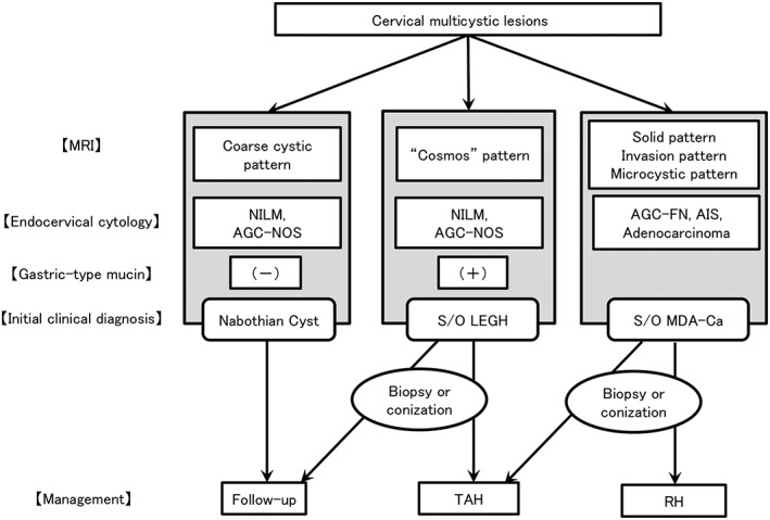
Figure 1.

Flow chart for the diagnosis and management of cervical multicystic lesions. This figure is a modified version of our original protocol. AGC‐FN, atypical glandular cells – favor neoplastic; AGC‐NOS, atypical glandular cells – not otherwise significant; AIS, adenocarcinoma in situ; Ca, carcinoma; LEGH, lobular endocervical glandular hyperplasia; MDA, minimal deviation adenocarcinoma; MRI, magnetic resonance imaging; NILM, negative for intraepithelial lesion or malignancy; RH, radical hysterectomy; S/O, suspicion of; TAH, total abdominal hysterectomy.
