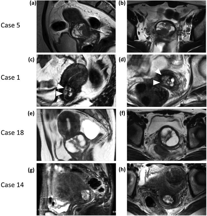
Figure 3.

Magnetic resonance imaging findings of patients who underwent hysterectomy, as shown in Table 2. (a,b) A typical solid pattern was observed in case 5. (c,d) A combination of solid pattern and invasion pattern. Microcystic and solid components (arrows) existed in the lateral portion of the cervix, suggesting stromal invasion, as observed in Case 1. (e,f) A typical cosmos pattern observed in Case 18 (i.e., small cysts or solid parts were surrounded by larger cysts). (g,h) A microcystic pattern: the aggregation of micro cysts, with the absence of large surrounding cysts or signs of invasion in Case 14.
