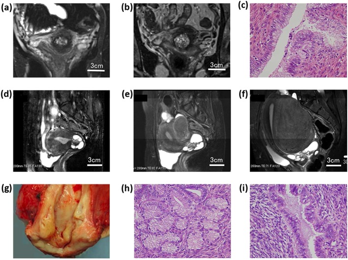
Figure 4.

Magnetic resonance imaging (MRI) and pathologic findings of two cases with increased lesion sizes during the follow‐up. (a–c) Case 21. (a) MRI showed the lesion size to be 15 × 10 × 8 mm at the first visit. (b) The lesion increased to 21 × 21 × 14 mm with more small cysts. (c) Hysterectomy specimen histologically showed lobular endocervical glandular hyperplasia (LEGH) with atypia. (d–i) Case 22. (d) The lesion size was 39 × 33 × 33 mm with a typical cosmos pattern at the first visit. (e) The lesion size increased 4 years later. (f) The lesion increased 66 × 45 × 37 mm 12 years after the first MRI. (g) A hysterectomy specimen showing a watery discharge. (h,i) LEGH with atypia was noted.
