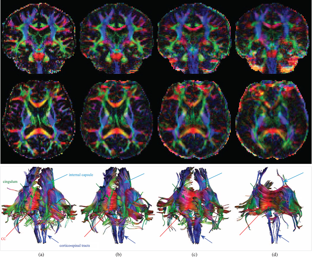
Figure 4.
Color-coded FA and tractography results comparison in a volunteer subject (2–2). Coronal (top) and transverse (middle) planes along with tracts passing CC. Cingulum tracts are completely missing in VVR and Orig. The FA and tracts obtained from MT-SVR are much more similar to those of the GS compared to the ones obtained from VVR and Orig. (a) gold standard (GS) (b) MT-SVR (c) VVR (d) Orig.

