Figure 1.
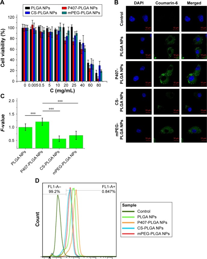
Cytotoxicity and cellular uptake of different surface-modified PLGA NPs in L929 cells.
Notes: (A) Cytotoxicity of NPs after incubation for 24 h as determined by CCK-8 assay. Percent viability of cells was expressed relative to the control cells. Results are shown as mean ± SD (n=6). (B, C) Confocal fluorescent microscopy and semi-quantitative fluorescence analysis showing the cellular uptake of different surface-modified, coumarin-6-loaded NPs after coincubation for 1 h (***P<0.001). Scales in the figure represent 10 µm. F-value is the ratio of MFI in each cell of various PLGA NPs to that of unmodified PLGA NPs. Results are shown as mean ± SD (n=30). (D) Cellular uptake of surface-modified coumarin-6-loaded NPs after coincubation for 1 h detected by flow cytometry.
Abbreviations: CCK-8, cell counting kit-8; CS, chitosan; DAPI, 4′,6-diamidino-2-phenylindole; MFI, mean fluorescence intensity; mPEG, methoxy poly(ethylene glycol); NPs, nanoparticles; P407, poloxamer 407; PLGA, poly(lactic/glycolic acid); SD, standard deviation; h, hour.

