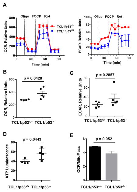
Figure 4. Effect of loss of p53 on mitochondria activity in leukemic splenocytes of TCL1 mice.
A, Basal oxygen consumption rate (OCR) and extracellular acidification rate (ECAR) was measured inleukemic splenocytes isolated from TCL1/p53+/+ mice (n=4) and TCL1/p53−/−mice (n=5). Three measurement cycles in duplicate was taken for each mouse sample. Representative graphs for OCR and ECAR are shown. Dotted lines indicate the time points of injection of 1 μM oligomycin (Oligo), 4 μM FCCP, or 2μM Rotenone (Rot). B, Comparison of the mean basal OCR between TCL1/p53+/+ mice (n=4) and TCL1/p53−/− mice (n=5). C, Comparison of the mean ECAR between TCL1/p53+/+ mice (n=4) and TCL1/p53−/− mice (n=5). D, Cellular ATP levels in the splenocytes from TCL1/p53+/+ and TCL1/p53−/− mice (n=4). E, Comparison between TCL1/p53+/+ mice (n=4) and TCL1/p53−/− mice (n=5) for their relative OCR normalized by mitochondrial mass measured by MitoTracker Green staining.

