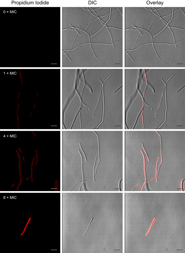
Figure 4.
Fluorescence microscopy reveals the uptake of propidium iodide (PI) by B. subtilis ATCC 47096 after treatment with JBIR-100. Cells were exposed at mid-log phase to vehicle alone (DMSO), 1× (8 µM), 4× (32 µM), or 8× (64 µM) MIC of JBIR-100, which was followed by washing and treatment with PI. Left panels, fluorescence of PI; middle panels, differential interference contrast (DIC); right panel, overlay of PI and DIC channels.

