Fig. 1.
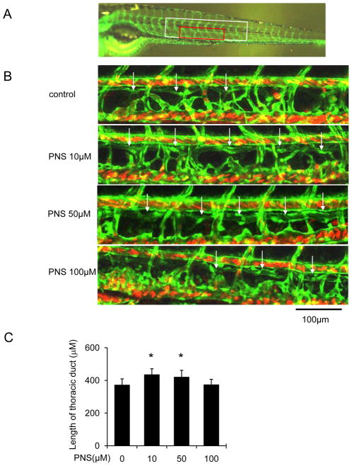
The lymphatic thoracic duct formation of zebrafish was increased by PNS in concentration dependent manner. The 48 hpf zeberafish (fli1:egfp; gata1:dsred) was treated with different concentrations of PNS (10, 50, 100μM) for 48 hours. Embryos treated with 0.2% DMSO served as a vehicle control. (A) Confocal image of the 96 hpf. zebrafish (fli1:egfp) vascular system. White boxed region indicates ten segment of thoracic duct length for quantitation in C; Red boxed region shows approximate location of regions imaged in B. (B) Representative confocal images show that PNS increased lymphatic thoracic duct formation of zeberafish, white arrow indicates lymphatic thoracic duct, and white star indicates lack of lymphatic vessel. Scale bars, 100 μm. (C) Quantitation of the length of lymphatic thoracic duct. Values are mean ± SD of 9–11 zeberafishes. *P<0.05 vs. vehicle control group.

