Figure 1.

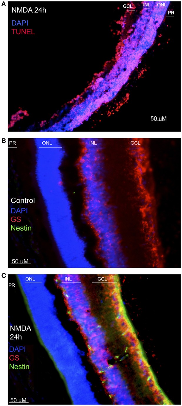
Effect of NMDA injury. (A) TUNEL test performed on a damaged retina, 24 h after injury. Cell nuclei are shown in blue, and apoptotic cells in red. (B) Immunofluorescence against nestin and glutamine synthase (GS) on an intact retina. MG, positive to GS, is shown in red. No nestin-positive cells are detected. (C) Immunofluorescence against the same markers on a damaged retina, extracted 24 h after injury. Co-labeling of GS (red) and nestin (green) indicates that MG are expressing the progenitor-associated marker as a response to damage. (ONL, outer nuclear layer; INL, inner nuclear layer; GCL, ganglion cell layer; PR, photoreceptor layer). Calibration bar: 50 μm.
